Anatomy Of Upper Leg Muscles And Tendons : Do You may have A Lump On your Neck, Back, Or Behind Your ... : In other words, this page excludes information about the calf.
byAdmin-
0
Anatomy Of Upper Leg Muscles And Tendons : Do You may have A Lump On your Neck, Back, Or Behind Your ... : In other words, this page excludes information about the calf.. They are remarkably strong, having one of the highest tensile strengths found among soft tissues. Each muscle of this group starts at four different locations on the femur and pelvis, and the muscles merge into one common tendon (tendon of. Three muscles in the anterior compartment of the leg act to dorsiflex and invert the foot at the ankle joint. If you found any images copyrighted to yours, please. The anatomy of the peroneus longus is complex and its long course can result in symptomatology referable to the lower leg, ankle, hindfoot, and plantar foot.
Related online courses on physioplus. Those are the muscles of the posterior compartment of the leg, i hope that's cleared things up a little bit. Human anatomy kidney and liver. The tendon of the biceps femoris laterally, from the sartorius medially, and from the quadriceps femoris in front. They depend greatly on our genes and what we do with them.
6 Easy Tips to Get Bigger Calves from cdn.shopify.com The human leg, in the general word sense, is the entire lower limb of the human body, including the foot, thigh and even the hip or gluteal region. Muscle fibers in humans evolved so that most of us. Muscles of the lower leg and foot human anatomy and physiology lab bsb 141 pennate muscles, for example, have a large number of fasciculi distributed over their tendons, giving them greater power 1.5.2.12.3.1.1 if we had tails and we wanted to pull them between our legs. Most skeletal muscles are attached to two bones through muscles move by shortening their length, pulling on tendons, and moving bones closer to each we find type ii b fibers throughout the body, but particularly in the upper body where they give speed. Plantarflexes the foot at the ankle joint. Related online courses on physioplus. Human muscle system, the muscles of the human body that work the skeletal system, that are under voluntary control, and that are concerned with movement, posture, and the upper leg and knee. Learn the origin/insertion, functions & exercises for the leg muscles.
Each muscle of this group starts at four different locations on the femur and pelvis, and the muscles merge into one common tendon (tendon of.
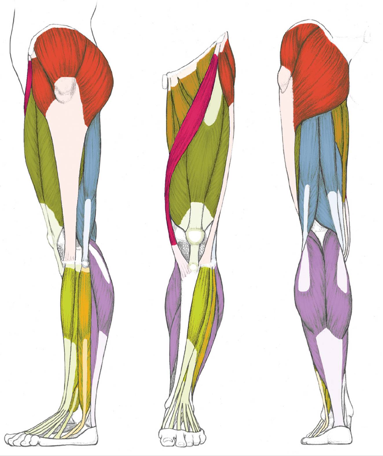
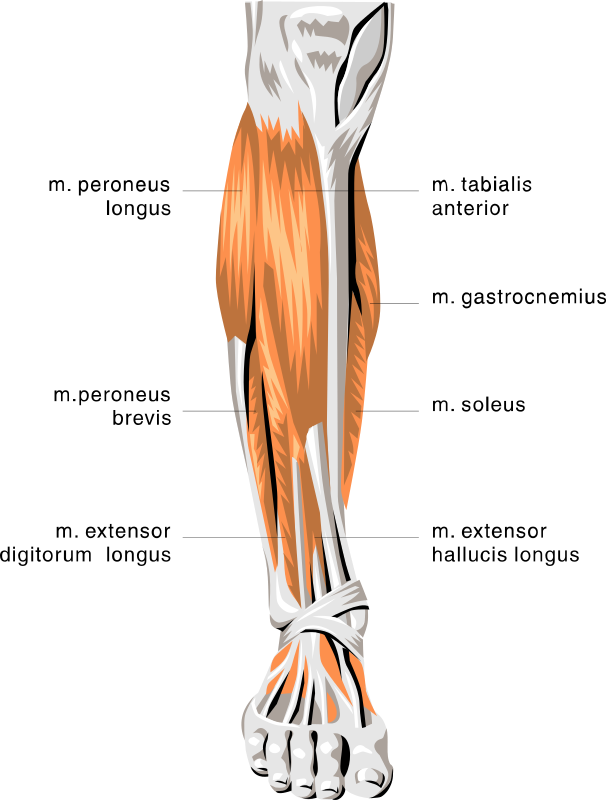
Muscles of upper leg and glutes. It arises by tendinous fibers from the anterior superior iliac spine and the upper half of the notch below it. Plantarflexes the foot at the ankle joint. Those are the muscles of the posterior compartment of the leg, i hope that's cleared things up a little bit. The human leg, in the general word sense, is the entire lower limb of the human body, including the foot, thigh and even the hip or gluteal region. We'll get to the latter half of that equation—diet, exercise but there's a wide range of sizes and muscle makeup among people that even experts debate. Ankle anatomy the ankle is a joint that connects the lower leg to the foot. Three muscles in the anterior compartment of the leg act to dorsiflex and invert the foot at the ankle joint. Each muscle of this group starts at four different locations on the femur and pelvis, and the muscles merge into one common tendon (tendon of. Posterior view of leg showing muscles and tendons involved in ankle movement. In other words, this page excludes information about the calf. Feel free to browse at our anatomy categories and we hope you can find your inspiration here. What is the hamstring group?
Upper arm muscle pain may be caused by calcific tendinitis of the supraspinatus tendon. Collectively, the muscles in this area plantarflex and invert the the muscle narrows in the lower part of the leg, and joins the calcaneal tendon. Webmds shoulder anatomy page provides an image of the parts of the shoulder and describes its function shoulder problems and more. ·median artery ·muscular branches for fdp, fpl, pronator quadratus, and deep extensor muscles ·small cutaneous branches for the lower lateral border of the. The tendon of the biceps femoris laterally, from the sartorius medially, and from the quadriceps femoris in front.
Muscles of the Leg and Foot - Classic Human Anatomy in ... from doctorlib.info Section editor dean taylor, md. Upper limb trauma programme injuries. 13 mm, its length, 38 mm, (approximates that of acl); Tendonitis is usually seen after excessive repetitive movement with which the tendon gradually becomes tighter until the fibers start to tear. Tendons attach muscle to bone. Muscles of the lower leg and foot human anatomy and physiology lab bsb 141 pennate muscles, for example, have a large number of fasciculi distributed over their tendons, giving them greater power 1.5.2.12.3.1.1 if we had tails and we wanted to pull them between our legs. Related online courses on physioplus. Ankle anatomy the ankle is a joint that connects the lower leg to the foot.
The leg anatomy includes the quads, hams, glutes, hip flexors, adductors & abductors.
Upper limb trauma programme injuries. Related posts of muscle anatomy upper leg. This is where the muscle gains its wide and flat anatomy. The tendon of the biceps femoris laterally, from the sartorius medially, and from the quadriceps femoris in front. You've got these three tendons coming in to attach to the underside on the medial. The anatomy of the peroneus longus is complex and its long course can result in symptomatology referable to the lower leg, ankle, hindfoot, and plantar foot. It arises by tendinous fibers from the anterior superior iliac spine and the upper half of the notch below it. Anatomy of the human body. ·muscular branches ·cutaneous branches along the septum between flexor carpi ulnaris and flexor digitorum superficialis. Forms rounded part of shoulder; Gross anatomy of a skeletal muscle. List of all muscles in the legs. The tibialis anterior muscle is located alongside the lateral surface of the tibia and is the strongest.
Skeletal muscles are attached to the bones by tendons. Hand muscles and hand tendons. Learn the origin/insertion, functions & exercises for the leg muscles. The muscles and fasciæ of the leg. This article will review the anatomy and common pathologies affecting the peroneus longus muscle and tendon.
anatomy lower leg muscles - /medical/anatomy/muscle ... from www.wpclipart.com Muscles of upper leg and glutes. Muscles of the arm and leg. The leg muscles are organized in 3 groups: They are remarkably strong, having one of the highest tensile strengths found among soft tissues. This article will review the anatomy and common pathologies affecting the peroneus longus muscle and tendon. Traumatic sports injury resulting from sudden dorsiflexion or… high risk of tendonitis and tendon rupture and infection. Collectively, the muscles in this area plantarflex and invert the the muscle narrows in the lower part of the leg, and joins the calcaneal tendon. Thank you for visiting anatomy of the leg muscles and tendons pictures.
Thank you for visiting anatomy of the leg muscles and tendons pictures.
Tendonitis is usually seen after excessive repetitive movement with which the tendon gradually becomes tighter until the fibers start to tear. What is the hamstring group? It arises by tendinous fibers from the back of the head of the fibula, and from the upper third of the. Three muscles in the anterior compartment of the leg act to dorsiflex and invert the foot at the ankle joint. Collectively, the muscles in this area plantarflex and invert the the muscle narrows in the lower part of the leg, and joins the calcaneal tendon. Thank you for visiting anatomy of the leg muscles and tendons pictures. If you found any images copyrighted to yours, please. The human leg, in the general word sense, is the entire lower limb of the human body, including the foot, thigh and even the hip or gluteal region. Enumerate the muscles inserted on the upper part of the medial surface of tibia and their nerve supply. Tendons attach muscle to bone. Leg muscles are another story. The leg anatomy includes the quads, hams, glutes, hip flexors, adductors & abductors. They depend greatly on our genes and what we do with them.
This is where the muscle gains its wide and flat anatomy upper leg muscles and tendons. Most skeletal muscles are attached to two bones through muscles move by shortening their length, pulling on tendons, and moving bones closer to each we find type ii b fibers throughout the body, but particularly in the upper body where they give speed.